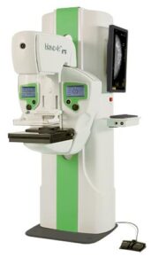
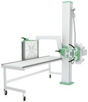
“MAMMO 4 MT PLUS” X-RAY SCREENING MAMMOGRAPHY DEVICE
The main advantages of the mammograph are ease of operation and reliability, high resolution, low radiation dose of the patient and staff, automatic selection of exposure parameters, high quality of the mammograms obtained.
The high performance of the device allows you to increase the capacity of the department, which is a significant advantage for screening studies.
Screening mammograph “Mammo-4MT-Plus” is supplied with a digital flat-panel receiver for indirect conversion of 24×30 cm format. The receiver provides high-speed acquisition of high-quality digital images. The technology of indirect conversion of X-ray radiation into an electrical signal and specially developed software guarantee the stability of operation and the receipt of images of the highest quality, regardless of the ambient temperature.
Full-format digital receiver
To obtain X-ray images, a specially designed high-resolution digital X-ray receiver is used.
The X-ray laboratory’s ARM allows you to fully automate the process, simplifying the work and minimizing the time of the study. The system transmits the received diagnostic images to the doctor’s ARM station and the PACS system according to the standard DICOM 3.0 protocol, and also receives lists of assigned studies (DICOM work lists).
Doctor’s ARM
We recommend completing the mammograph with an automated radiologist’s workplace (specialization: mammography), equipped with two specialized medical high-contrast monitors with a resolution of at least 5 Megapixels, providing the doctor with a full set of necessary tools for a full analysis of diagnostic images. The doctor’s workplace automates the work of describing images, preparing conclusions, printing X-ray images, as well as preparing a CD with images in DICOM format with software for viewing them on the computers of clinicians and patients.

